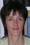
Prof. Dr. Anne SENTENAC
Anne SENTENAC, PhD, Directeur de Recherche au CNRS
Image: PrivateAnne Sentenac, Institut Fresnel, Marseille, is visiting the Faculty of Physics and Astronomy as an Abbe School of Photonics Visiting Professor in August 2012. During her stay she will give three lectures. On top of that, she will also give talks about some interesting research advances in the field of microscopy. She obtained her PhD in physics (electromagnetism) in 1993. Since 2001 she holds a permanent researcher position at the French National Research Center (CNRS), at the university of Marseille. Dr. Sentenac is a specialist in the field of electromagnetism and optics. She has published 77 articles and many novel and promising, mostly theoretical, ideas. Her current research activities include:
- propagation in random media (scattering by rough surfaces, rough inhomogeneous films and new formulations for the effective permittivity of random media)
- optical components with resonant gratings (development of narrow-band filters and directional sources with dielectric waveguide gratings. Modification of the dispersion relation of guided waves with periodic structures)
- high resolution far-field optical imaging (development of an optical diffraction tomograph which presents a better power of resolution than classical microscopes)
- numerical methods (volume integral method for simulating the field diffracted by a 3D object in a complex environment (multilayer, grating))
website of Anne SentenacExternal link
Lecture 1: Improving the Axial Resolution of Optical Microscopes Using a Mirror for Holding the Sample Instead of a Glass Slide
Time: August 23, 2011, 10:00
Place: Lecture Hall 2, Helmholtzweg 5, 07743 Jena
The resolution of optical microscopes is always much better in the plane of observation than in the depth of the sample. This unfortunate feature limits the ability of optical microscopy for performing three-dimensional imaging with high resolution. The poor in-depth resolution comes from the fact that, in a microscope, the sample is illuminated and observed from only ONE side. Indeed, if the sample was illuminated and observed from all possible angles (as in a scanner), the resolution would trivially be the same in all directions. In this talk, we present a possible solution to address this problem. It consists in replacing the usual glass slide on which the sample is laid by a mirror. Thanks to the mirror, the sample is illuminated and observed from both sides. Now, the difficult part is to separate the top and bottom views of the sample.
Lecture 2: Imaging Non-Fluorescent Objects with Optical Diffraction Tomography
Time: August 24, 2011, 10:00
Place: Lecture Hall 2, Helmholtzweg 5, 07743 Jena
Optical diffraction tomography is a recent microscopy technique that consists in recording many holograms of an object under different angles of illumination. The set of holograms is then injected in a reconstrfction algorithm to yield the quantitative map of permittivity of the object. In this talk, we show the interest of this technique for obtaining 3D highly resolved images of the object. We show that a resolution much better than the Rayleigh or Abbe limit can be obtained under certain conditions.
Lectures 3 and 4: Basics of Imaging with Electromagnetic Wave Probing
Time: August 6, 2012, 10:00, and August 8, 2012, 15:00
Place: Lecture Hall, Helmholtzweg 4, 07743 Jena
In these two lectures, we will first study the interaction between light and a (non-fluorescent) sample. We will see how the scattered field is related to the permittivity of the object. Then, we will show how this relationship can be used to image the object with electromagnetic wave probing and will study the resolution limits of this approach. Finally, we will present different experimental implementations of this principle (in the microwave, optical and X-ray domains).
Lecture 5: Study of the Classical Widefield Microscope (non-fluorescent sample)
Time: August 20, 2012, 10:00
Place: Lecture Hall, Helmholtzweg 4, 07743 Jena
In this course, we will explain how the image obtained in a classical widefield microscopy is linked to the sample. Starting from the relationship between the scattered field and the sample (seen in the first course), we will build an expression relating the recorded intensity to the permittivity of the sample. We will show that this approach can be applied to other types of microscopes (phase microscopy, dark-field). This course is inspired from the paper by Streibl, JOSA-A, 1985. To attend this course, it is strongly advised to attend the 2 preceeding lectures (first course "basics of imaging with electromagnetic wave probing").
